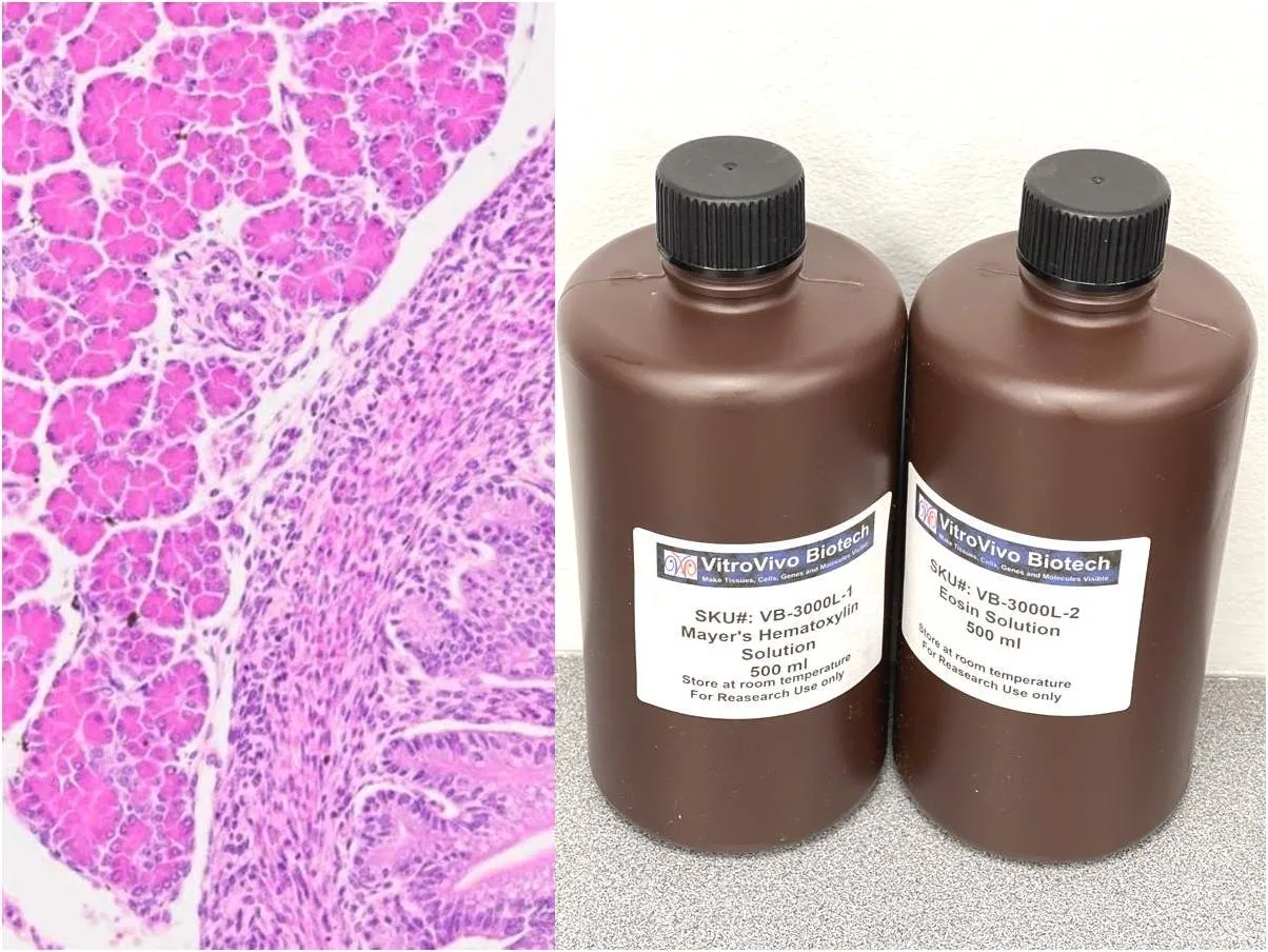
Routine histology often works best as a staged process rather than a single staining decision. A hematoxylin and eosin staining kit gives researchers a broad view of nuclei, cytoplasm, extracellular material, and tissue architecture before more targeted methods are introduced.
That first overview can reveal whether the section contains the expected anatomy, whether morphology is preserved, and which regions deserve closer examination. Once those questions are answered, a special stain can be selected to investigate a more specific tissue component.
Start With a Reliable Morphology Baseline
H&E staining is useful because it creates clear contrast across many tissue types. Hematoxylin highlights nuclei in blue tones, while eosin stains much of the cytoplasm and extracellular material pink to red, allowing researchers to evaluate overall organization.
When abnormal extracellular deposits are suspected in a research sample, congo red staining amyloid provides a more focused approach. Congo Red binds beta-pleated sheet structures associated with amyloid fibrils, helping investigators localize deposits that may not be confidently characterized on routine morphology alone.
Use Routine Staining as a Map
A general stain can guide where additional analysis should occur. Researchers can identify vessels, neural regions, connective tissue, inflammatory areas, or other anatomical landmarks before deciding which sections or regions need specialized evaluation.
This is especially useful when valuable tissue is limited. Instead of applying several special stains without a clear plan, investigators can review an H&E section first and reserve adjacent sections for methods that answer specific follow-up questions.
Understand What Congo Red Adds
Congo Red provides information that H&E does not. In suitable tissue sections, amyloid deposits appear red, while a hematoxylin counterstain provides blue nuclear contrast that helps preserve the anatomical context of the signal.
The method can be used with formalin-fixed, paraffin-embedded tissue and may also be applied to frozen sections. Section preparation still matters, because thickness, fixation, differentiation, dehydration, and mounting can influence the final appearance.
Plan Adjacent Sections Carefully
Using neighboring sections can connect general morphology with targeted staining without consuming unnecessary tissue. One slide may establish the overall architecture, while the next is used to examine a suspected deposit or structure in greater detail.
Researchers should remember that adjacent sections are similar but not identical. Small deposits may change from one cutting level to the next, so orientation, section order, and anatomical landmarks should be recorded during the study.
Control Technical Variation
Staining consistency is essential when several samples will be compared. Fixation time, section thickness, deparaffinization, reagent exposure, differentiation, and washing can all change the visual result even when the underlying biology is similar.
Standard operating procedures help reduce this variation. Running control tissue alongside experimental sections also provides a reference for expected staining performance and makes troubleshooting easier when a batch looks unusual.
Think Beyond Color Alone
A red deposit should be interpreted in its morphological setting rather than treated as an isolated observation. Researchers should consider location, shape, distribution, surrounding tissue, and whether the staining pattern is consistent across related sections.
The same principle applies to H&E. Strong color is not the goal by itself; useful contrast should make structures easier to distinguish while preserving enough detail for meaningful comparison between regions and experimental groups.
Standardize Imaging and Review
Microscope settings can affect how both routine and special stains appear in captured images. Magnification, illumination, exposure, white balance, and digital processing should remain consistent when slides are being compared.
If scoring or image analysis is planned, criteria should be defined in advance. Researchers may assess deposit area, distribution, regional involvement, or another parameter, but the same rules should be applied across the complete sample set.
Use Archived Tissue Efficiently
FFPE tissue can support multiple rounds of sectioning and staining, making it valuable for studies that evolve. A thoughtful slide-allocation plan can reserve material for routine histology, special stains, immunostaining, and possible future assays.
Good records also improve reproducibility. Documenting block identity, section level, staining run, control tissue, and imaging conditions creates a clear history that helps researchers interpret results and repeat the workflow when necessary.
Conclusion
Routine morphology and targeted histochemistry serve different but complementary purposes. A broad tissue overview can identify where an abnormal feature is located, while a specialized method can provide stronger evidence about the nature of that feature.
By planning section use, standardizing staining, including controls, and interpreting targeted signals within their anatomical context, researchers can obtain more information from limited tissue while keeping each observation connected to the wider experimental question.